
LEARNING FROM IMAGES – Dysfunctional Dialysis Catheter
Tunneled dialysis catheter is a frequently used vascular access to initiate hemodialysis treatment for patients with end stage renal disease. Right internal jugular vein is the preferred site due to its straight continuation in to the superior vena cava [1]. A well positioned tunneled catheter will have a smooth transition from the tunnel in to the internal jugular vein. Improper placement technique can lead to early dysfunction and inadequate dialysis treatment. Fig. (1A) shows an acute kink in the tunneled portion of the catheter resulting in catheter dysfunction. The ideal treatment option, when possible, is to create a new tunnel utilizing the same venous puncture thus preserving the venous access site. Additionally, the procedure is short with minimal discomfort to the patient. Fig. (1B) shows a new catheter with a smooth curve at the transition point from the subcutaneous tunnel into the internal jugular vein.
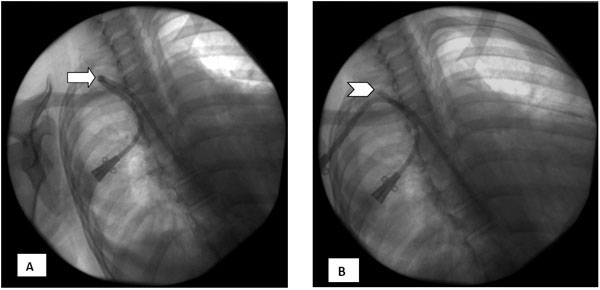
(A) A right internal jugular vein tunneled dialysis catheter with a kink (marked by arrow). (B) Well positioned tunneled dialysis catheter utilizing the same venous puncture site. Old catheter has been cut at the venous puncture site (marked by chevron).
CONFLICT OF INTEREST
The author confirms that this article content has no conflict of interest.
ACKNOWLEDGEMENT
Declared none.

