
LEARNING FROM IMAGES Seroma Related to Prosthetic Arteriovenous Graft
INTRODUCTION
Seroma is a complication unique to prosthetic arteriovenous graft (AVG), resulting as a consequence of transudation of sterile and clear serum, which is confined by a non-secretory fibrous pseudomembrane [1]. Although the precise etiology remains elusive, it is thought to be related to failure of the surrounding connective tissue to incorporate the graft [2]. The incidence of 2-4% reported in literature may not be a true representation due to frequently seen spontaneous resolution of seroma [2]. A seroma is typically located close to the arterial anastomosis and usually occurs within the first month after placement of the AVG [3] (Fig. 1). Ultrasound-guided needle aspiration of the fluid yielding serous or gelatinous material can be of both diagnostic and therapeutic significance. When in doubt, the fluid should be submitted for white blood cell count and bacterial evaluation. Treatment options include serial aspirations, surgical evacuation with or without drain placement, graft wrapping with microfibrillar collagen and surgical excision of the entire seroma with tissue debridement while leaving the graft in situ [1]. However, recurrence is not uncommon and a total graft excision and placement of a new conduit may rarely be necessary [1]. Although, it is a non-infectious process initially, repeated intervention can lead to secondary infection and access loss. Besides unsightliness, if left untreated, it can lead to wound infection, skin necrosis, graft thrombosis, or loss of the available cannulation area [2].
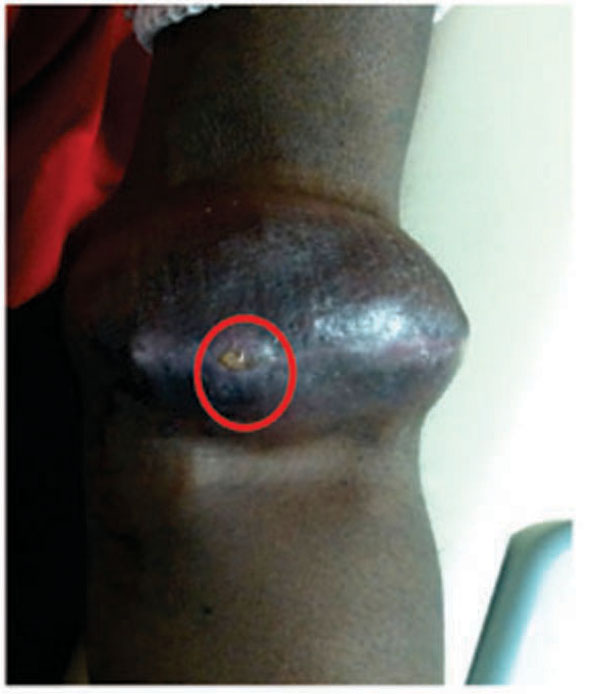
An end stage renal disease patient with a left forearm brachio-basilic arteriovenous graft who developed a golf ball size seroma 3-weeks after graft placement. Note a clear serous discharge upon gentle compression (marked by red circle).
CONFLICT OF INTEREST
The authors confirm that this article content has no conflict of interest.
ACKNOWLEDGEMENTS
Declared none.

